
LNP vs Electroporation for CRISPR Delivery in HSCs: Key Insights for Researchers
CRISPR-Cas9 gene editing has become a key technology in therapeutic development, making precise genome modification possible in areas such as immuno-oncology, rare diseases, and regenerative medicine. Its success depends on efficient delivery of Cas9 mRNA and sgRNA into cells, especially hematopoietic stem cells (HSCs), which are difficult to transfect and highly sensitive to stress.
Application
Lipid Nanoparticles
Industry
Biopharma
Key Products
RESEARCH SUMMARY
Are LNPs a viable alternative to electroporation for CRISPR delivery in HSCs?
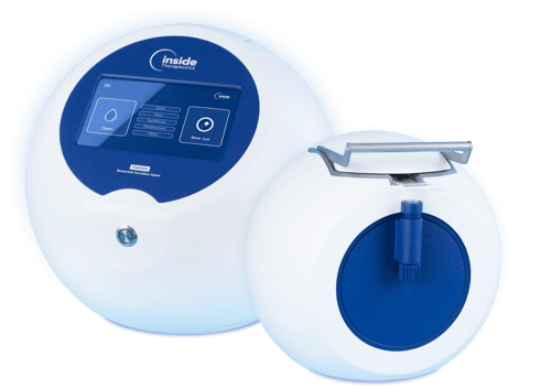
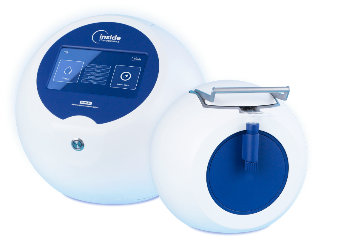
This article highlights key findings from a study conducted by Inside Therapeutics in collaboration with the BIGRes research team, evaluating lipid nanoparticle (LNP) delivery versus electroporation for eGFP mRNA and CRISPR-Cas9 delivery in hematopoietic stem cells (HSCs). The original work was performed using the TAMARA® microfluidic formulation platform.
The study investigated whether LNPs could provide a viable alternative to electroporation, a widely used transfection method that is often associated with reduced cell viability and limitations for clinical translation.
Access the original application note from Inside Therapeutics.
BACKGROUND
Why is Exploration of LNPs for CRISPR delivery Important?
Hematopoietic stem cells are among the most challenging cell types to transfect successfully. Their importance in cell and gene therapy applications means researchers need delivery technologies that achieve high editing efficiency while preserving cell viability and functionality.
Traditionally, electroporation has been the preferred approach. However, increasing interest in RNA therapeutics and scalable manufacturing has driven the exploration of lipid nanoparticles as a gentler alternative for both research and future therapeutic applications.
KEY FINDINGS
How do LNPs compare with electroporation for CRISPR delivery in HSCs?
-
High RNA Delivery Efficiency: The study demonstrated that LNPs formulated using the TAMARA® platform achieved near-complete eGFP mRNA transfection across all tested doses in HSCs. Flow cytometry analysis showed almost 100% eGFP-positive cells while maintaining excellent viability.
-
Effective CRISPR-Cas9 Gene Editing: When delivering Cas9 mRNA and sgRNA targeting the β2-microglobulin (B2M) gene, LNP formulations achieved knockout efficiencies approaching 100% at optimal doses. Editing performance was comparable to electroporation.
-
Superior Cell Viability: One of the most significant findings was the impact on cell health. While electroporation resulted in approximately 20% cell loss, LNP-mediated delivery maintained cell viability at around 99%. This could be an important consideration for workflows where cell recovery is critical.
-
Improved Reagent Efficiency: The researchers also observed that LNP-mediated delivery achieved similar editing outcomes while requiring substantially less sgRNA than electroporation, potentially supporting lower reagent costs and improved process scalability.
Why Microfluidic Formulation Matters
While the study focused on biological outcomes, it also highlights the importance of reproducible nanoparticle formulation.
Microfluidic mixing technologies enable precise control over particle formation, helping researchers generate LNPs with consistent size, encapsulation efficiency, and performance characteristics. The study's LNPs were formulated using the TAMARA® microfluidic platform, producing particles close to 100 nm in diameter, a range commonly associated with efficient intracellular delivery.
For organizations developing RNA therapeutics, consistency during formulation is often as important as the nucleic acid payload itself, particularly when moving from early-stage discovery towards scale-up and manufacturing.
CONSIDERATIONS
Implications for Cell and Gene Therapy Development
The results suggest that LNP-mediated delivery has the potential to offer several advantages over traditional electroporation workflows:
-
Improved cell viability while maintaining high transfection efficiency in difficult-to-transfect cells.
-
Compatibility with future in vivo delivery strategies.
-
Improved scalability through controlled microfluidic nanoparticle production.
These characteristics make LNP technology an increasingly important area of interest for organizations working on next-generation gene-editing therapies.
About This Research
This article is intended as a summary and interpretation of research performed by Gregory Noel, Robin Oliveres and Michel Cogné in collaboration with Inside Therapeutics and the BIGRes team. The original data, conclusions and application note remain the work of the original authors.
Get your free copy.
Open this application note in a downloadable format.
FAQs
Low-volume formulation allows researchers to evaluate more experimental conditions while conserving valuable materials. This can be particularly important when working with limited quantities of CRISPR payloads during optimization studies.
Encapsulation efficiency measures how much of the payload is successfully incorporated into the lipid nanoparticle. Higher encapsulation efficiency can help maximize the use of valuable materials and improve formulation development workflows.
Reproducibility helps researchers determine whether observed differences are linked to formulation design rather than process variation. Consistent nanoparticle production can support more reliable optimization and screening studies.
Microfluidic technology is widely used for CRISPR-LNP formulation because it provides accurate control over nanoparticle assembly through the controlled mixing of lipid and aqueous streams under laminar flow. These low-shear conditions help preserve the integrity of nucleic acids while enabling the production of uniform nanoparticles with optimized size, polydispersity index (PDI), and encapsulation efficiency. As a result, microfluidics improves formulation consistency, scalability, and overall CRISPR delivery performance.
Researchers often seek formulation platforms that support both early-stage screening and larger batch volumes. Using a consistent formulation approach across development stages can simplify optimization and technology transfer workflows.
Researchers can reduce material waste during CRISPR-LNP formulation screening by using low-volume workflows that require smaller amounts of lipids, nucleic acids, and other valuable reagents. Advanced microfluidic platforms such as TAMARA further minimize waste by using compressed air to purge the system at the end of each run, ensuring that residual sample is recovered rather than left in the tubing. This maximizes material utilization, lowers development costs, and helps conserve precious research materials.
Improving CRISPR delivery efficiency typically requires optimization of both the payload and delivery system. Researchers often investigate factors such as particle size, encapsulation efficiency, lipid composition and formulation conditions to maximize delivery performance and generate reproducible results. LNP-based delivery systems are increasingly being explored as an alternative to viral approaches across a range of gene-editing applications.
Variability in formulation processes can affect particle size, encapsulation efficiency and overall nanoparticle quality. Researchers often use controlled formulation methods, such as microfluidics, to improve consistency and reduce experimental variability during optimization studies.
Encapsulation efficiency can be influenced by formulation parameters including lipid composition, flow rates, mixing conditions and nucleic acid concentration. Optimizing these variables helps researchers improve payload utilization and formulation performance.
There is no fixed number. Researchers typically investigate multiple formulation conditions before advancing lead candidates. Low-volume screening approaches can support broader formulation exploration while conserving valuable materials.
Researchers often address this challenge through low-volume screening and highly efficient formulation workflows. This enables more experimental conditions to be evaluated from the same quantity of payload, supporting informed optimization decisions.
FURTHER RESOURCES
You may also be interested in these pages:
TAMARA RNA-LNP Formulation System
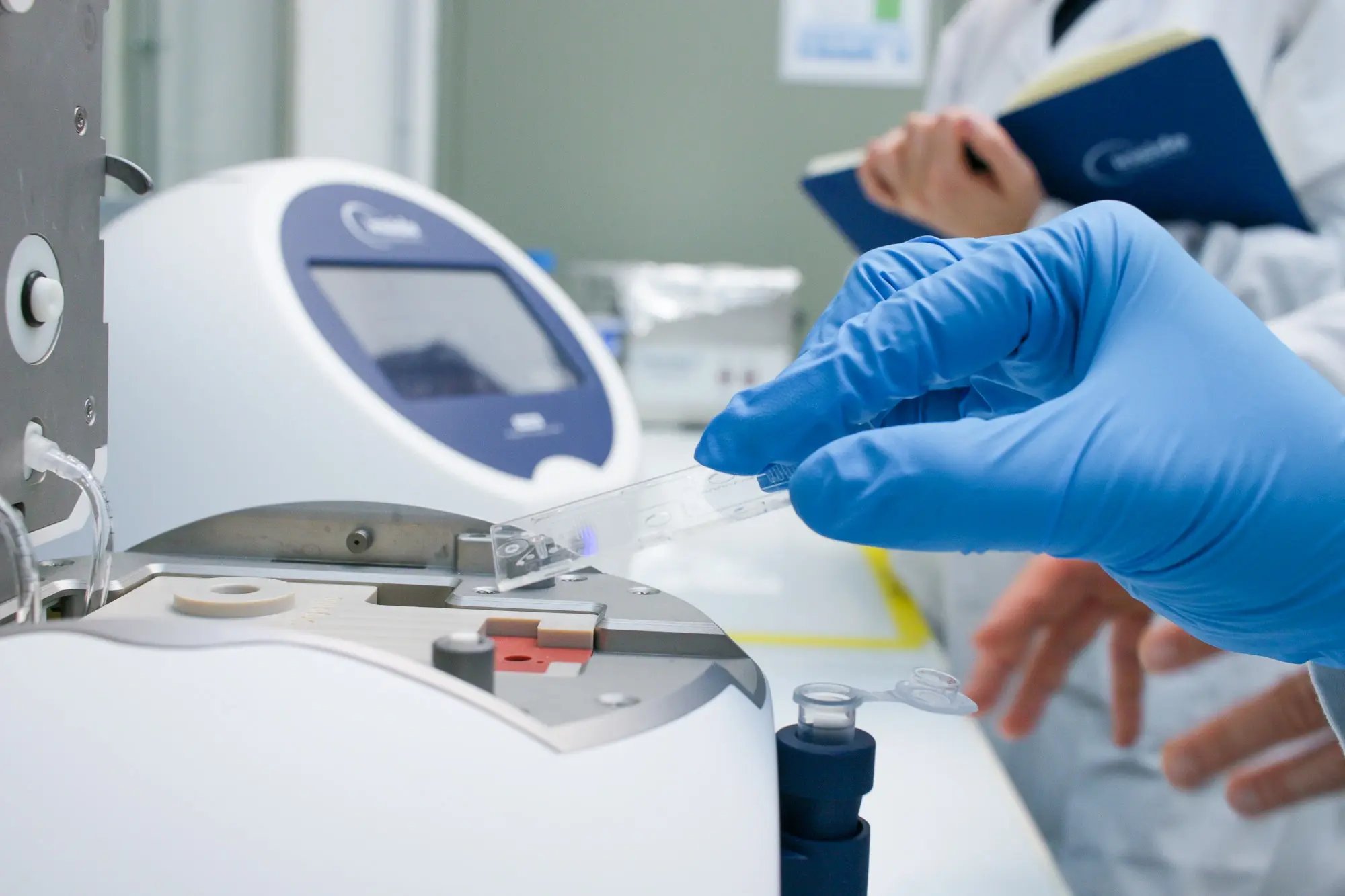
A simple to use benchtop machine for formulating lipid nanoparticles, covering low volumes for screening up to in-vivo scale production.
.webp?width=800&height=533&name=TAMARA-Labo-Fevrier-2025-InsideTx-27%20(1).webp) Explore
Explore
CRISPR Drug Delivery Using LNPs
The critical role of delivery in CRISPR research and how lipid nanoparticles are revolutionizing gene editing therapies.
 Explore
TAMARA Specifications & Resources
In-depth details, unboxing, standard protocols and specifications for the TAMARA nanoparticle formulation system.
 Explore