

 Fast
Fast
Processing
 High
High
Yields
 Improved
Filtration
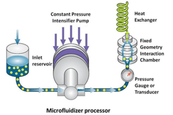
Figure 1: Schematic of Microfluidizer® technology
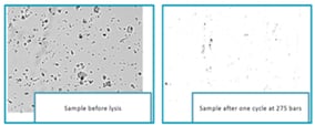
Figure 2: Microscopic images of cells before and after the Microfluidizer® technology homogenization step
 Figure 3: Amount of AAV recovered after cell lysing with freeze-thaw method & Microfluidizer® technology
APPLICATION
How can Microfluidizer® technology be used in cell lysis for gene therapy?
This case study compares virus recovery efficiency using the freeze-thaw method and Microfluidizer® technology. In the study, AAV vectors were produced in HEK293 cells and resuspended in Tris buffer before cell lysis with each method. For freeze-thaw processing, the cell suspension went through five cycles of freezing in a dry ice/isopropanol bath followed by thawing at 37°C to rupture the cells. For Microfluidizer® processing, the cell suspension was passed once through a Microfluidizer® processor fitted with an H30Z (200 µm) Interaction Chamber™ at 4,000 psi (275 bar). In both workflows, the lysate was centrifuged for 30 minutes to remove debris and then analyzed by droplet digital polymerase chain reaction (ddPCR) to measure AAV titer. As shown in Figure 2, microscopic images taken before and after Microfluidizer® processing indicate that a single pass was sufficient to rupture almost all cells.
Figure 3 shows that Microfluidizer® technology delivered approximately 50% higher AAV recovery than the freeze-thaw method. It also reduced processing time significantly. The freeze-thaw workflow required 5 hours to complete, while the full Microfluidizer® process took less than 1 hour, including preparation and sanitizing steps.
SUMMARY
Benefits of Microfluidizer® technology in cell lysing for gene therapy?
In summary, Microfluidizer® technology offers several practical benefits for viral vector production. It supports high yield by rupturing cells effectively while helping protect viral vectors. It may also reduce issues such as virus agglomeration and membrane binding, both of which can lower vector recovery.
The technology is scalable from laboratory development to full production, and biopharmaceutical models are available for cGMP-compliant manufacturing. It can also simplify downstream processing by breaking cells gently but efficiently, producing larger cell wall fragments that are easier to separate from smaller viral vectors. In addition, the process can be adjusted to shear cellular DNA and reduce viscosity, which helps make clarification and filtration easier. Because no chemical lysis method is required, there is no added detergent removal step, downstream processing can be simpler, and the need for costly nucleases such as Benzonase can be reduced or eliminated.
.webp?width=900&height=450&name=Microfluidizer-Technology-Scale-Up-1%20(1).webp)
Microfluidizer® Technology for Cell Disruption
See how the choice of cell disruption method affects key properties such as particle size, disruption efficiency, and protein release.
.webp?width=400&height=400&name=Microfluidizer%C2%AE%20Technology%20%20for%20Cell%20Disruption%20(1).webp) Explore
Explore
Blog: What is Cell Disruption and What are the Methods?
Learn more about the process of breaking cells open (otherwise known as lysing) in order to obtain the intracellular fluid.
 Explore
How it Works: Microfluidizer® Technology
Microfluidizer® Technology delivers superior results by combining a constant pressure pumping system with a unique fixed-geometry Interaction Chamber™.
 Explore